|
Common Conditions Treated and Services Offered
Periodontal Disease
What is it? Disease affecting the tissues and bones that support the teeth. We offer a few different options to treat periodontal disease. These are extraction of the teeth, gingivectomy/gingivoplasty to eliminate pseudo-pocketing, root planing (closed and open), and guided tissue regeneration with the use of bone grafts.
Fractured or Non-Vital Teeth
We offer endodontic therapy as an alternative to tooth extraction. This is a great option for fractured or non-vital canine and carnassial teeth. The pulp is removed and the tooth is filled with an inert filling material, allowing the patient to maintain the important chewing and grasping functions of these teeth.
Before |

After |

Dental Radiograph Following Root Canal Therapy
Malocclusions
Endodontics can also be used to treat malocclusions (when one or more teeth are in the incorrect position). This becomes a problem when the soft tissues are traumatized from this misplacement.
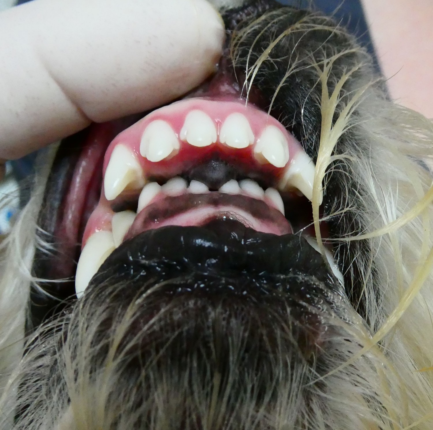 
Before
Crown reduction followed by vital pulp therapy or root canal therapy is an option for treating traumatic malocclusion. This allows us to reduce the height of the teeth causing trauma while maintaining their function.

After
The application of an orthodontic appliance is an option for some malocclusion treatment plans. Slow, constant pressure of the appliance guides the teeth into a more appropriate position. The images below demonstrate the use of restoration extensions to adjust the alignment of the canine teeth.
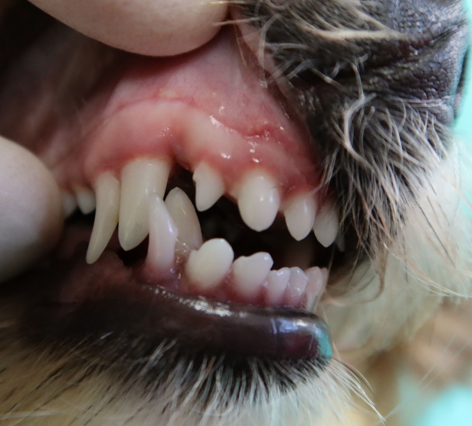 
Before

Midway
(after removal of deciduous teeth and extension application)

After
Retained Deciduous Teeth
Deciduous (baby) teeth should fall out by approximately 6 months of age. Retained deciduous teeth can cause malocclusion and/or crowding, which can lead to early periodontal disease. These teeth should always be extracted.
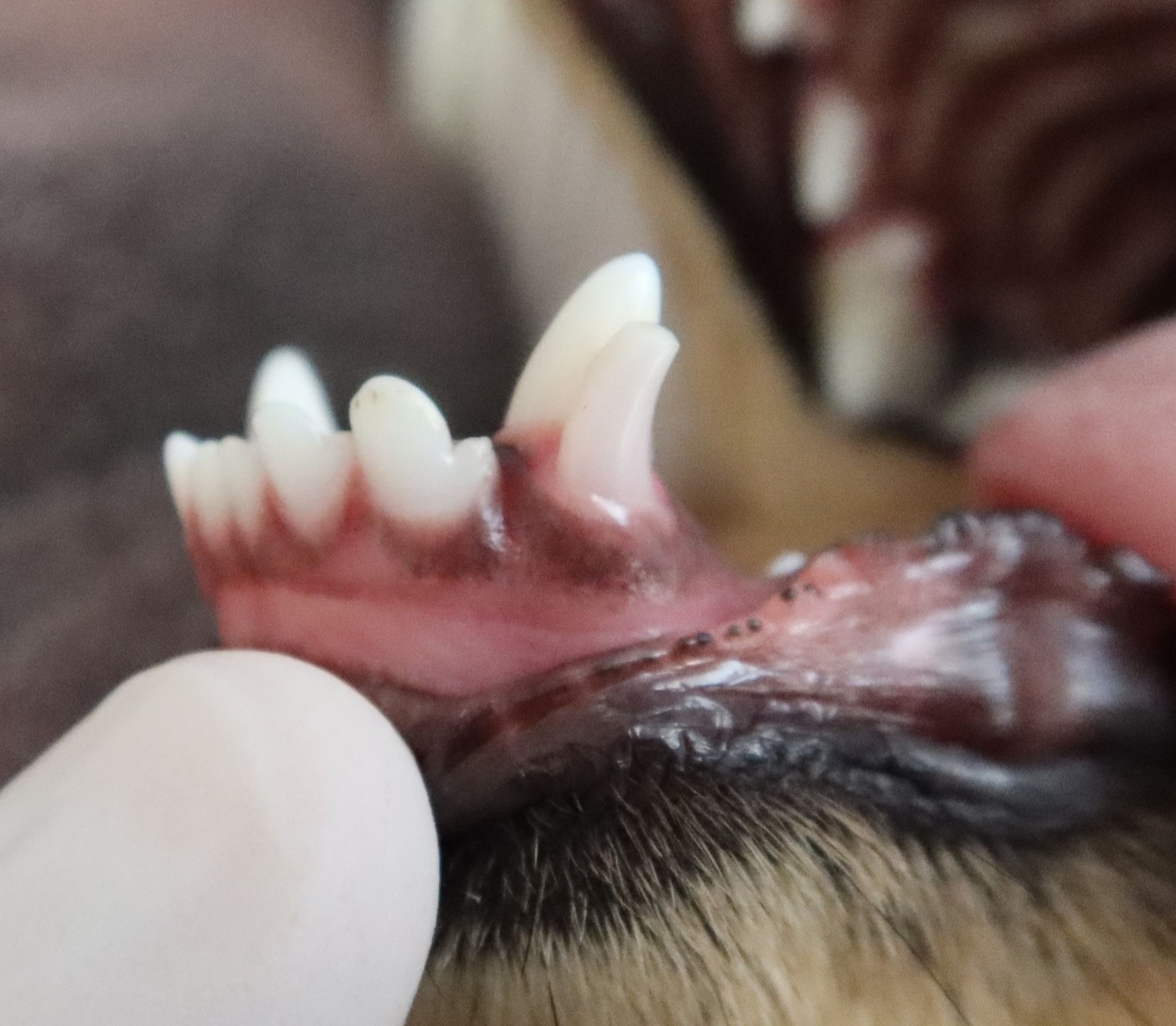 
Before

After
Pyogenic Granulomas
Pyogenic granulomas can form in cats with malocclusions. This is caused by the upper carnassial tooth contacting the soft tissue on the lower mandible. Often, this causes a painful lesion and also periodontal disease.
 
Oral Tumors
We diagnose and treat oral tumors through biopsy and surgical excision. Examples of frequently seen oral tumors include Melanoma, Squamous Cell Carcinoma, Fibrosarcoma, Peripheral Odontogenic Fibroma, Acanthomatous Ameloblastoma, Osteosarcoma, and Papillary Squamous Cell Carcinoma.
Not all oral tumors are metastatic, however some benign tumors can still be locally invasive, displacing teeth, getting in the way when chewing, and making life uncomfortable for the patient. These tumors can be removed with the appropriate margins and the patient can go on with a great quality of life. We also offer staging, surgical excision and counseling for the treatment of metastatic oral disease.
In some patients, to achieve clean margins, Mandibulectomy (removal of a portion of the lower jaw) or Maxillectomy (removal of a portion of the upper jaw) may be necessary.
More often than not, there isn't a drastic change noticeable to the patient's face structure.

Before |

After |
Unerupted Teeth
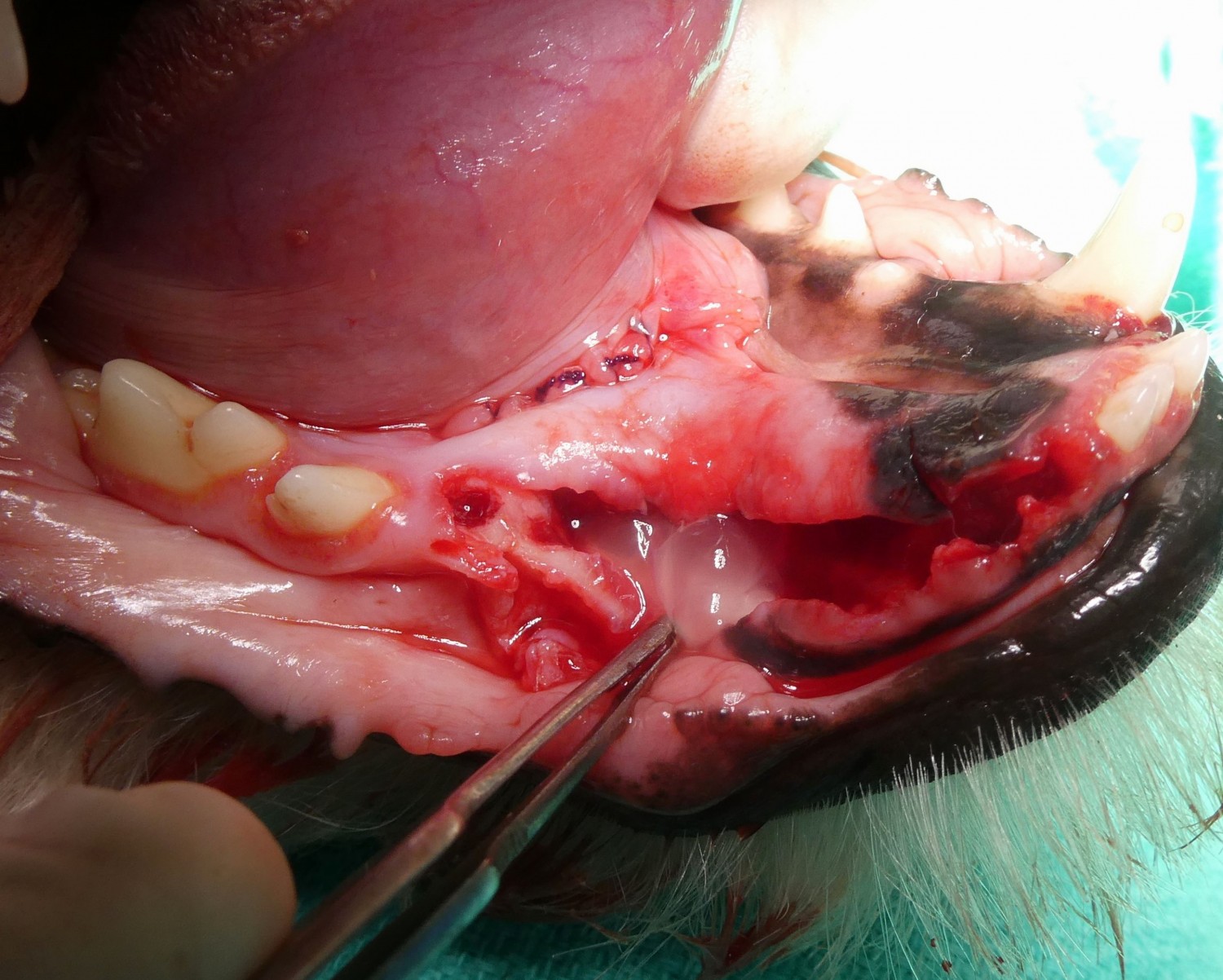
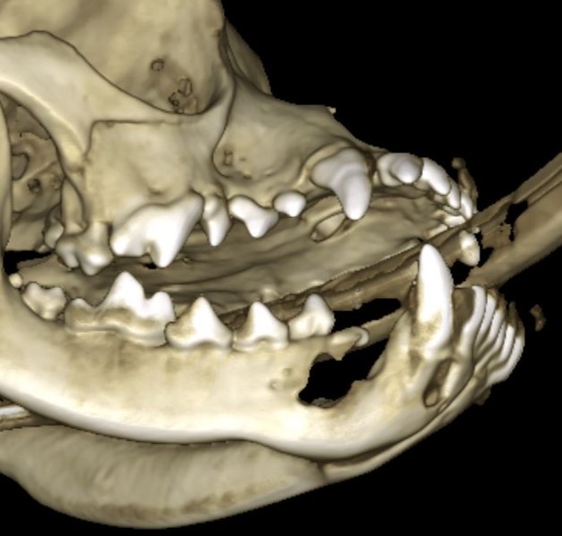
If a tooth is missing, imaging should be done to determine if that tooth never developed or if the tooth is present but unerupted. Unerupted teeth can cause expansile cysts to develop. These teeth should always be extracted.

Clinical Exam |

Cone Beam CT Scan
|

Dental Radiograph |
Odontogenic Cysts
Untreated odontogenic cysts can cause bone destruction and displacement of teeth. Treatment includes the complete removal of the cyst lining, any unerupted teeth associated with the cyst, and extraction of teeth that have extensive bone loss due to expansion of the cyst.
Gingival Overgrowth
We have multiple options to aid in the treatment of gingival enlargement or periodontal pocketing. Gingivectomy/gingivoplasty, guided tissue regeneration (placement of a bone graft), root planing, and crown lengthening for incompletely erupted teeth.

Before gingivectomy/gingivoplasty |

After gingivectomy/gingivoplasty |

Before crown lengthening |

After crown lengthening |
Immune-mediated Diseases
We offer oral surgery and medical management for these cases. Feline Stomatitis and Canine Chronic Ulcerative Stomatitis (CCUS) are shown in the images below respectively.
|

Feline Stomatitis
|

Canine Chronic Ulcerative Stomatitis (CCUS)
|
Tooth Resorption
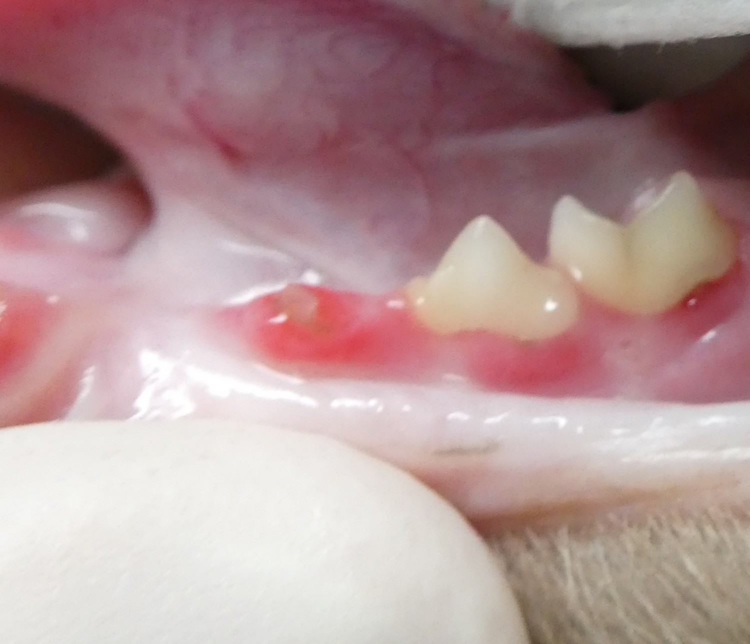
Teeth with resorptive lesions can be painful and difficult to extract. We have extensive experience in extracting these teeth. Unfortunately, extraction is the only acceptable treatment for teeth with this condition.
Prosthodontics
Crowns can be applied to non-vital (after root canal therapy) or vital strategic teeth. This is especially helpful in law enforcement dogs and pet dogs doing bite work. They can also be applied to feline teeth!

Dental Restorations
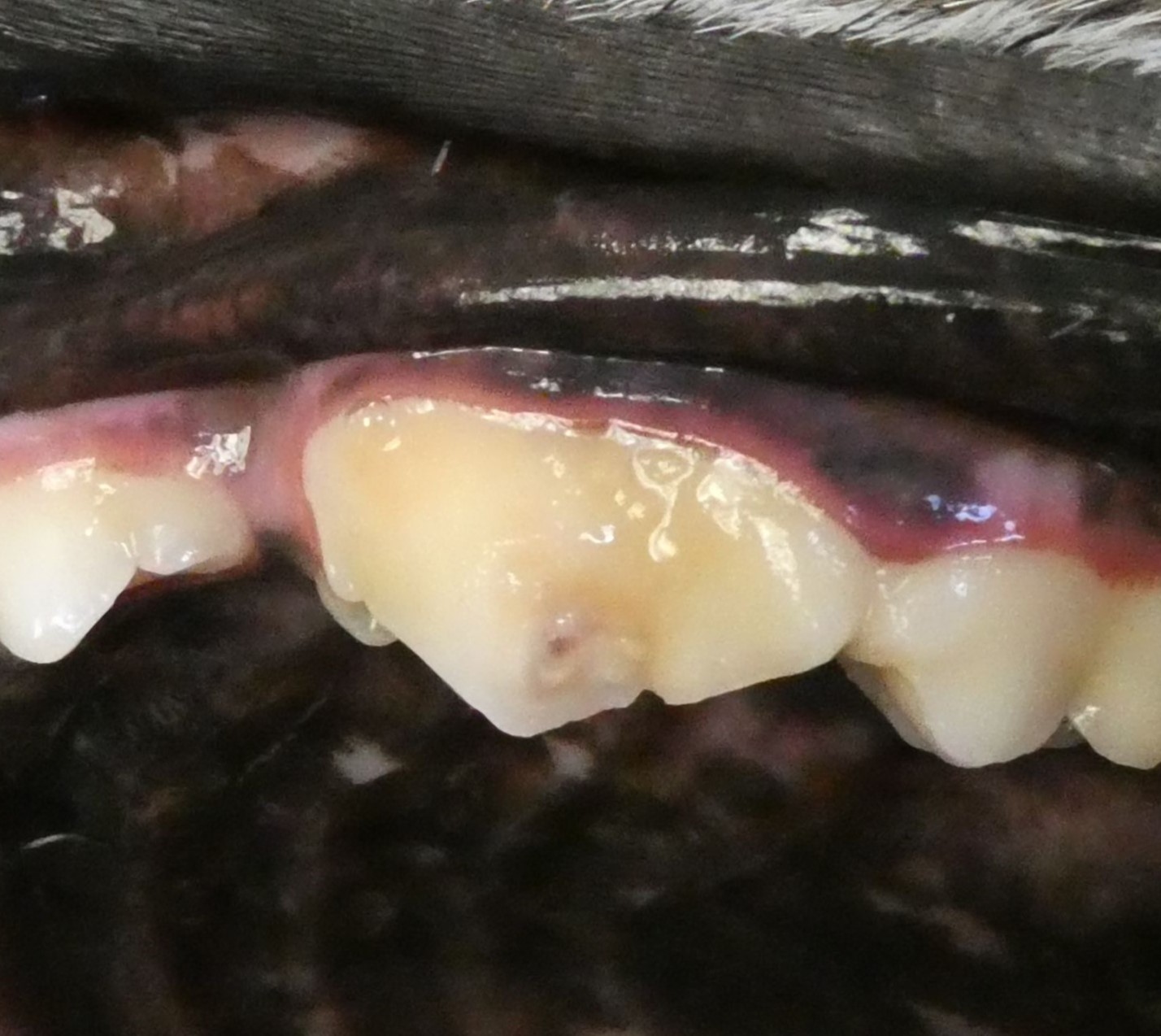
Caries
Dental caries (similar to cavities in humans) are uncommon in dogs, but can affect the teeth that are similar in shape to our teeth. The decay is removed and a composite filling is placed.

Before |

After |
Enamel Defects
Enamel defects can occur for a variety of reasons. Restorations can be applied to protect the porous dentin that is exposed.

Oro-nasal Fistula Repair
Oro-nasal fistulas (an abnormal communication between the mouth and the nasal cavity) can occur due to several different scenarios. Most commonly, they result following loss or extraction of an upper canine tooth. It is important that these are surgically repaired to prevent food and water from entering the nasal cavity, potentially leading to aspiration pneumonia.

Oro-nasal fistula |

After oro-nasal fistula repair |
Jaw Fracture Repair
Jaw fracture can occur from trauma or long standing periodontal disease. We offer repair and management of these cases using a variety of techniques to restore the patient’s original occlusion (interdigitation and arrangement of the upper and lower teeth).

Before |

With temporary wire placed |

Before |

With wire and composite splint |
|